
Cardiothoracic and Vascular Surgery
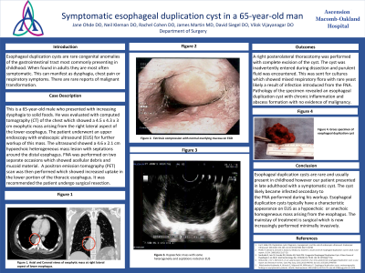
This is a 65-year-old male who presented with increasing dysphagia to solid foods. He was evaluated with computed tomography (CT) of the chest which showed a 4.5 x 4.3 x 3 cm exophytic mass arising from the right lateral aspect of the lower esophagus. The patient underwent an upper endoscopy with endoscopic ultrasound (EUS) for further workup of this mass. The ultrasound showed a 4.6 x 2.1 cm hypoechoic heterogeneous mass lesion with septations around the distal esophagus. FNA was performed which was nondiagnostic. A positron emission tomography (PET) scan was then performed which showed increased uptake in the lower portion of the thoracic esophagus. It was recommended the patient undergo surgical resection.

Jane Ohde, DO
Minimally Invasive Foregut Fellow
AHN Esophageal Institute
Pittsburgh, PA, US
Vilok G. Vijayanagar, DO
St. John Macomb-Oakland Hospital
Warren, MI, US
James Martin, MD
Chief of Cardiothoracic Surgery
Ascension Macomb-Oakland
Warren, MI, US
David Siegel, DO
General Surgeon, Attending, Program Director
Ascension Macomb-Oakland
Warren, MI, US
Neil Kleman, DO
Resident
Ascension Macomb-Oakland
Madison Heights, MI, US
Rachel Cohen, DO
Resident
Ascension Macomb-Oakland
Madison Heights, MI, US